Mind The Gap: Cryo-EM Reveals Lipids Intercalating Connexin Assemblies
Using single particle cryo-EM on plaque-mimicking nanodiscs, researchers determined multiple connexin assembly states and uncovered lipid-protein interactions that may help understand gap junction organization and function.
No Cell Is an Island
Multicellular organisms depend on constant communication between cells. Signals, nutrients, and electrical impulses flow through intricate cellular networks to coordinate everything from thought and movement to the maintenance of tissues and organs. While some messages travel over long distances through ligand-receptor signaling, others are exchanged through direct cell-cell contact. Gap junctions represent one of the most intimate forms of this communication: these membrane channels span two plasma membranes, physically connecting neighboring cells, allowing ions and small molecules to pass directly between them. These connections synchronize heart contractions, coordinate neuronal activity, and help tissues function as unified systems [1].
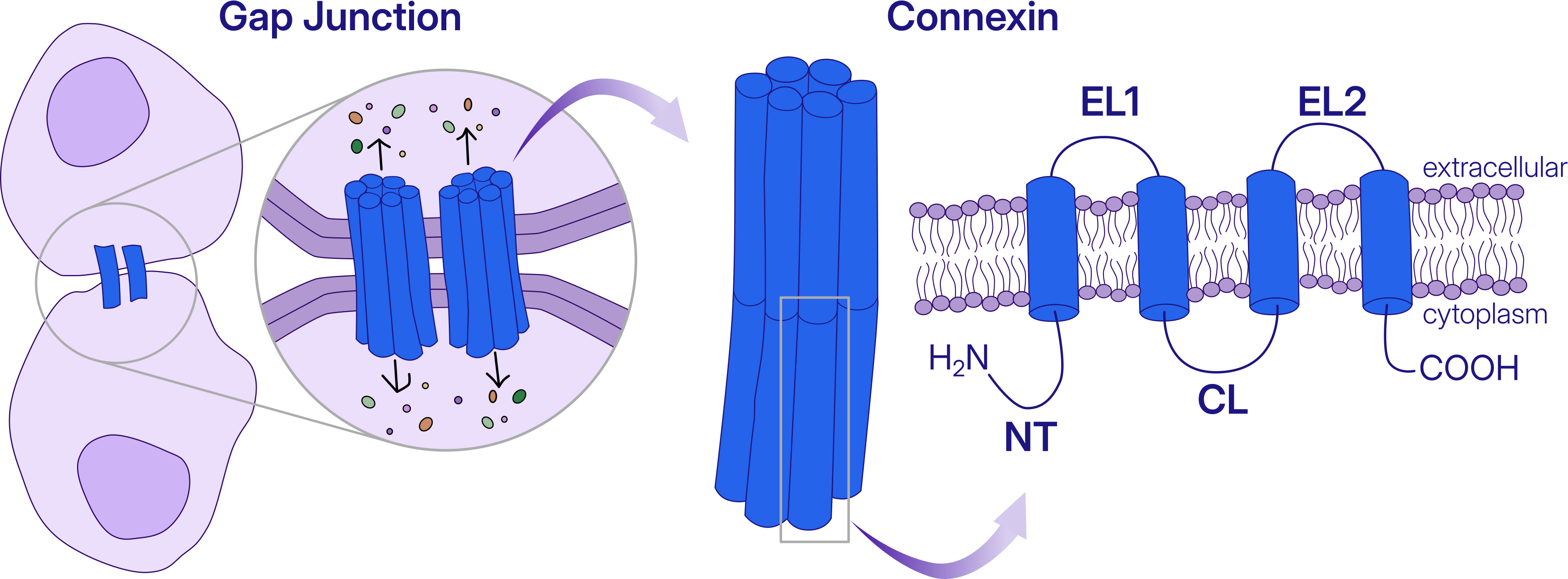
In a recent preprint, scientists at Oregon Health & Science University used cryo-EM to investigate native lens connexin-46/50 (Cx46/50), a major component of gap junctions. Connexin dysfunction is linked to a wide range of human diseases, including deafness, blindness, skin disorders, neuropathies, cardiac arrhythmias, and cancer [2].
No Protein Is an Island
Gap junction channels do not exist in isolation. Instead, they cluster into densely packed assemblies known as plaques, which can contain hundreds to thousands of channels arranged within the membrane. Studies of individual gap junction channels have revealed many fundamental features of connexin biology [3], [4], yet functional studies of intact plaques in tissues and model systems have revealed behaviors that cannot be fully explained by isolated channels alone [5].
“Gap junction plaques have been studied structurally for decades, but we have lacked a high-resolution view of how neighboring channels interact within the membrane.”
- Dr. Steve Reichow, Associate Professor, Oregon Health & Science University, (corresponding author)
To capture this higher-order organization, the authors reconstituted Cx46/50 into MSP-based lipid nanodiscs, incorporating multiple channels into miniature plaque-like complexes suitable for single particle cryo-EM analysis.
.](/images/blog/gap-junctions/blog-gap-junctions-figure2.png)
Even before data processing began, the sample hinted at the complexity ahead. Reconstitution produced a mixture of nanodiscs containing single channels, paired channels, and larger multi-channel assemblies. Rather than a uniform population, the researchers were faced with substantial structural heterogeneity, a challenge familiar to cryo-EM scientists, but also the key to understanding how plaque organization influences gap junction function.
Navigating Heterogeneity in Cryo-EM: Single- and Dual-Channel Connexins
The dataset was processed in CryoSPARC, where the central challenge quickly became clear: heterogeneity. While approximately 75% of the particles corresponded to single-channel assemblies - ultimately reconstructed to an impressive 1.8 Å resolution for comparison - the remaining 25% consisted largely of dual-channel assemblies. Unraveling the structural diversity within this population became one of the focus points of the analysis.
Micrograph denoising paired with template picking using a previously determined dual-channel structure was used for a first selection of the particles of interest. 2D Classification followed by multi-class Ab-Initio Reconstruction allowed further enrichment of the particles of the dual-channel. The authors then applied what they call “baited” Heterogeneous Refinement, seeded with two competing references: a single-channel map with D6 symmetry and a dual-channel map used during at template-picking stage. Two successive rounds of 3D Variability Analysis revealed that the dataset contained not only compositional heterogeneity (single versus dual channels) but also conformational heterogeneity within the dual-channel population itself.
.](/images/blog/gap-junctions/blog-gap-junctions-figure3.png)
The dual-channel assemblies adopted two distinct arrangements. In one, the channels were aligned around a centered tilt axis, producing equivalent cytosolic interfaces on both sides of the complex. In the second, the channels were offset relative to one another, creating asymmetric cytosolic interfaces. To avoid imposing incorrect symmetry during classification, symmetry was only applied at the final stage, resulting in a D2-symmetrized reconstruction for the centered assembly and a C2-symmetrized reconstruction for the offset assembly.
Assembly Formation Stabilizes Interface-Associated Lipids
While the overall architecture of individual connexin channels remained largely unchanged upon assembly formation, the dual-channel structures revealed a subtle but important difference.
“What surprised us most is that the channels do not appear to interact directly. Instead, lipids fill the space between them and help organize and provide plasticity to their high-order assembly. The 1.8 Å single-channel structure gave us a near-atomic open-state reference, allowing us to pinpoint lipid features that emerge specifically in the multi-channel assemblies. These findings suggest that the membrane itself helps build high-order plaques and shape how cells communicate with each other.”
- Dr. Steve Reichow, Associate Professor, Oregon Health & Science University, (corresponding author)
The N-terminal (NT) domain adopted essentially the same conformation in both single- and dual-channel structures, suggesting that channel clustering does not substantially alter this key gating element. However, the centered dual-channel structure revealed an interfacial lipids nestled between the two channels, with their headgroups directly engaging the open-state NT domain. Together with previous biochemical observations, this interaction supports the existence of a lipid-dependent gating mechanism.
This work is an example of what single-particle cryo-EM can reveal beyond the structure of an isolated protein. By recreating miniature gap junction plaques, the authors could determine multiple structures from the same heterogeneous sample at near-atomic resolution, making it possible to visualize lipid interactions that only emerge when channels assemble together.
The findings challenge the view of the membrane as a passive scaffold. Instead, they suggest that lipids actively stabilize connexin assemblies and may help tune how neighboring cells exchange signals. In other words, communication between cells is shaped not only by the proteins that form gap junctions, but also by the membrane environment that brings them together.
References
- Goodenough, D. A., & Paul, D. L. (2009). Gap junctions.Cold Spring Harbor Perspectives in Biology, 1(1), a002576. doi: 10.1101/cshperspect.a002576
- Delmar, M., Laird, D. W., Naus, C. C., Nielsen, M. S., Verselis, V. K., & White, T. W. (2018). Connexins and disease.Cold Spring Harbor Perspectives in Biology, 10(9), a029348. doi: 10.1101/cshperspect.a029348
- Kronengold, J., Srinivas, M., & Verselis, V. K. (2012). The N-terminal half of the connexin protein contains the core elements of the pore and voltage gates.The Journal of Membrane Biology, 245(8), 453-463. https://doi.org/10.1007/s00232-012-9457-z
- Yue, B., Haddad, B. G., Khan, U., Chen, H., Atalla, M., Zhang, Z., ... & Bai, D. (2021). Connexin 46 and connexin 50 gap junction channel properties are shaped by structural and dynamic features of their N-terminal domains.The Journal of Physiology, 599(13), 3313-3335. https://doi.org/10.1113/JP281339
- Bukauskas, F. F., Jordan, K., Bukauskiene, A., Bennett, M. V., Lampe, P. D., Laird, D. W., & Verselis, V. K. (2000). Clustering of connexin 43-enhanced green fluorescent protein gap junction channels and functional coupling in living cells. Proceedings of the National Academy of Sciences, 97(6), 2556-2561. https://doi.org/10.1073/pnas.050588497