Cryo-EM Reveals the Molecular Machinery of Tubulin Assembly
By capturing transient β-tubulin-bound intermediates with cryo-EM, researchers reconstructed the molecular mechanism of α/β-tubulin biogenesis, explaining how mutations in the assembly machinery lead to tubulin biogenesis disorders.
In the delicate process of brain and body development, many events must occur with extraordinary precision. Even minor disruptions to these processes, often beginning before birth, can have devastating consequences that become apparent during the first years of life. This is the case for genetic disorders such as infantile encephalopathy, corpus callosum hypoplasia, and Kenny-Caffey syndrome, all of which belong to a group of diseases known as tubulinopathies [1], [2], [3].
Tubulin is the main building block of microtubules, one of the major components of the cytoskeleton. These dynamic structures are essential for a wide range of cellular processes, including cell division, intracellular transport, maintaining cell shape, cilia function, neuronal migration, and axon growth [4]. Even a small fraction of defective tubulin has severe consequences. Whether caused by mutations in its building blocks (α- and β-tubulin) or by defects in the machinery that assembles them into functional heterodimers, the resulting tubulinopathies can affect the brain, eyes, peripheral nervous system, musculoskeletal system, and other organs. This translates to shorter life expectancy and a quality of life reduced by epilepsy, motor impairment, neurological disabilities and other physical complications [5].
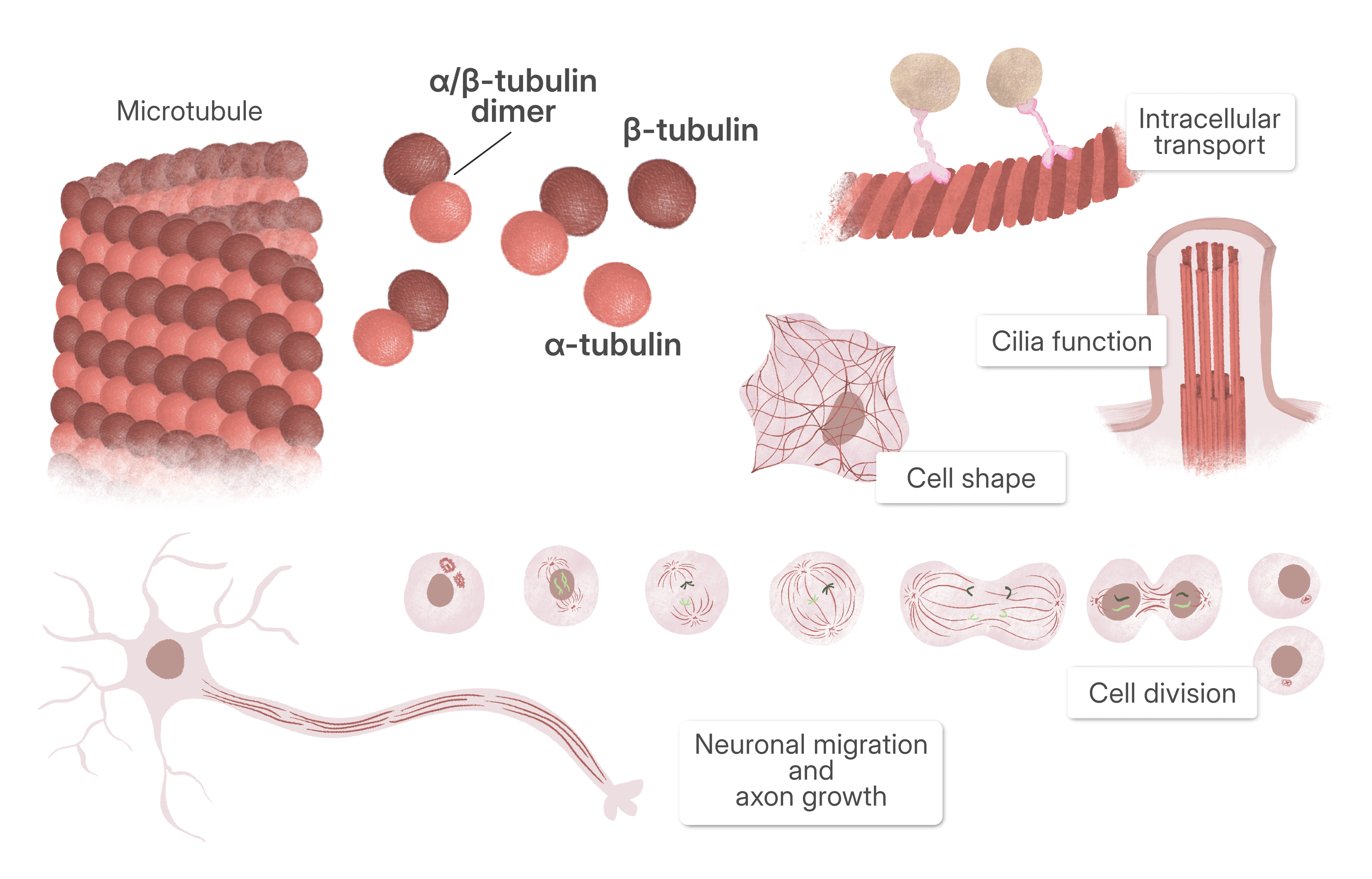
A growing subgroup of tubulinopathies arises from defects in tubulin biogenesis, the cellular pathway that assembles α- and β-tubulin into functional heterodimers. Rather than affecting tubulin itself, these disorders are caused by mutations in the tubulin cofactors and associated proteins, including TBCD, TBCE, and Arl2 [6].
The Challenge of Protein Reconstitution
Multiple mutations in the cofactors responsible for α/β-tubulin assembly have been identified, but despite their clinical importance, the molecular mechanism by which they assemble α/β-tubulin remained elusive. The assembly machinery is highly dynamic and involves multiple states, making it exceptionally difficult to study.
“For decades, the mechanism of α/β-tubulin dimer assembly was neglected, largely because the cofactor assemblies that regulate the process could not be reconstituted biochemically. More than a decade ago, our laboratory overcame this barrier by establishing recombinant reconstitution of the tubulin cofactor assemblies bound to tubulin in distinct functional states. Over the following decade, we refined this system to capture even more states, including the β-tubulin–bound cofactor assemblies.”
- Dr. Jawdat Al-Bassam, Associate Professor, UCDavis (corresponding author)
Recent cryo-EM studies performed at UCDavis have begun to reveal, at near-atomic resolution, how the TBC-DEG complex orchestrates tubulin assembly, providing the first molecular explanation for many of these diseases [7]. In a recent paper, published in Science Advances, researchers resolved the structures of the TBC-DE and TBC-DEG complexes in multiple functional states, capturing them bound to β-tubulin at different stages of the assembly pathway.
Two Sample Preparations, Three Datasets, Seven Cryo-EM Structures
From only two sample preparations scientists performed three cryo-EM data collections, ultimately generating seven distinct cryo-EM reconstructions, capturing previously unseen β-tubulin assembly intermediates.
“Cryo-EM was the key that unlocked these intermediates: advances in 3D classification and flexible refinement allowed us to resolve the mobile, transient elements within these complexes that had previously been inaccessible. Assembling this collection of structures ultimately enabled us to build a complete catalytic model for the process.”
- Dr. Jawdat Al-Bassam, Associate Professor, UCDavis (corresponding author)
Specifically, two different β-tubulin bound assemblies were reconstituted by co-expression: TBC-DE, composed of the tubulin cofactors TBCD and TBCE, and TBC-DEG, which additionally includes the small GTPase Arl2. To capture different oligomerization states, the TBC-DEG sample was imaged twice: once immediately after purification and once after mild crosslinking to stabilize dimeric intermediates.
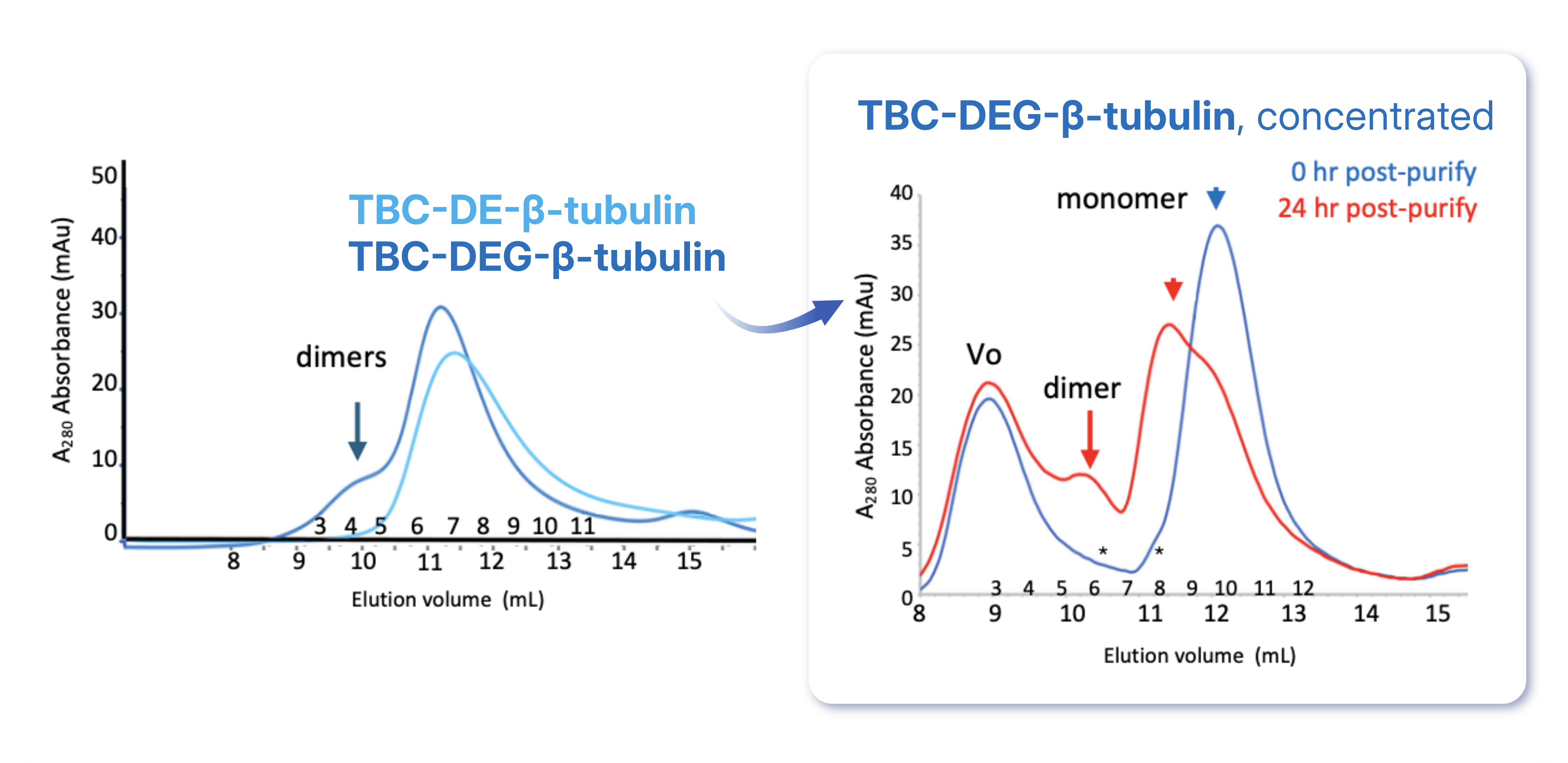
The paper describes four cryo-EM data processing workflows: one for the TBC-DE–β-tubulin monomer, one for the TBC-DE–β-tubulin dimer, one for the fresh TBC-DEG–β-tubulin monomer, and one for the crosslinked TBC-DEG–β-tubulin dimers. Although the overall processing strategy was similar, each workflow was tailored to the structural heterogeneity present in its respective dataset.
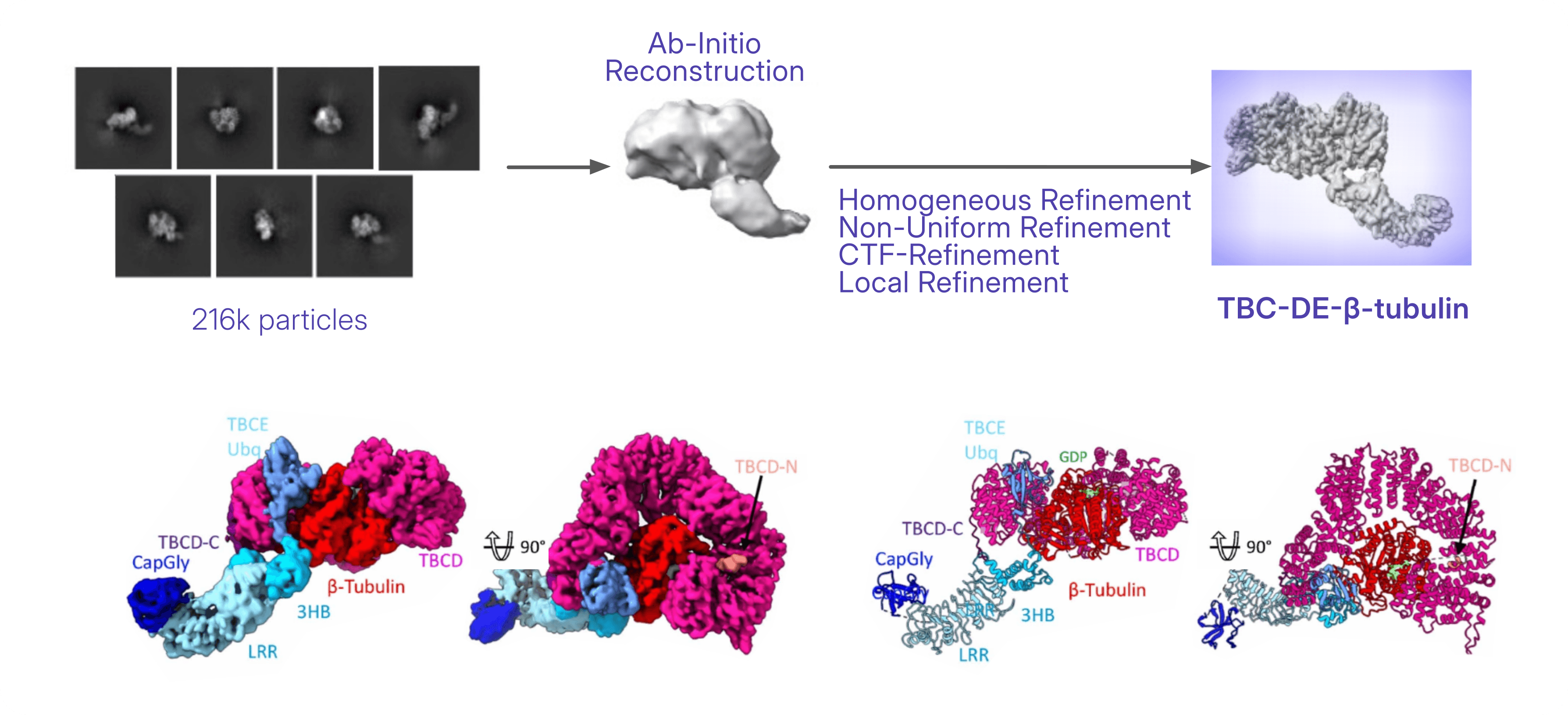
The TBC-DE–β-tubulin dataset contained both monomeric and dimeric particles. The monomeric structure was determined through Homogeneous Refinement, followed by Non-Uniform Refinement, and lastly CTF and Local Refinement, yielding the structure of the TBC-DE–β-tubulin complex. This represents the earliest experimentally observed intermediate in the assembly pathway, where β-tubulin is captured by TBCD and TBCE prior to Arl2 binding.
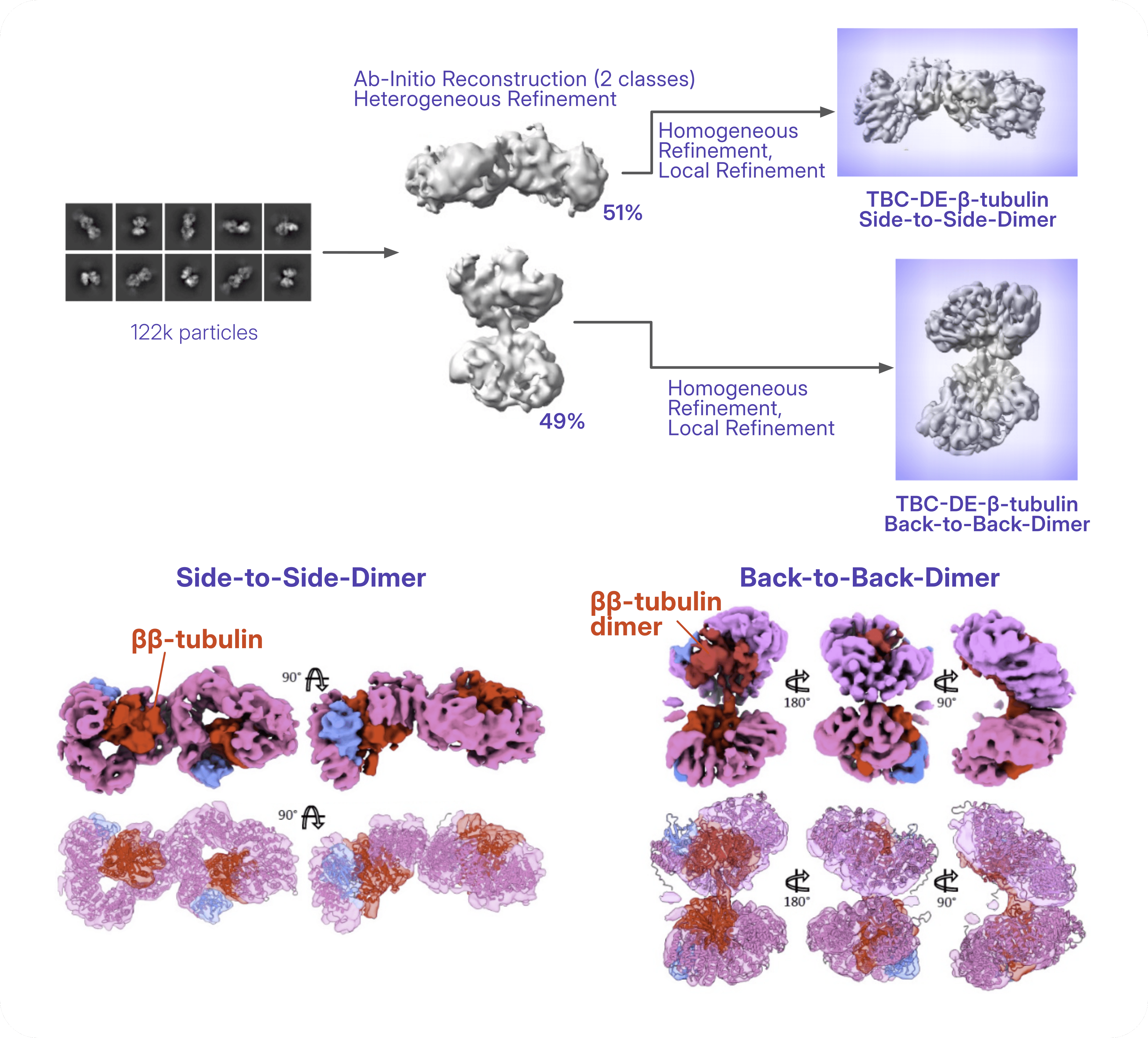
On the other side, the dimeric particles in the TBC-DE–β-tubulin dataset separated into two distinct assemblies (back-to-back and side-to-side dimer) using multi-class Ab-Initio Reconstruction followed by Heterogeneous Refinement in CryoSPARC. These dimeric assemblies are interpreted as alternative β-tubulin-bound oligomeric states.
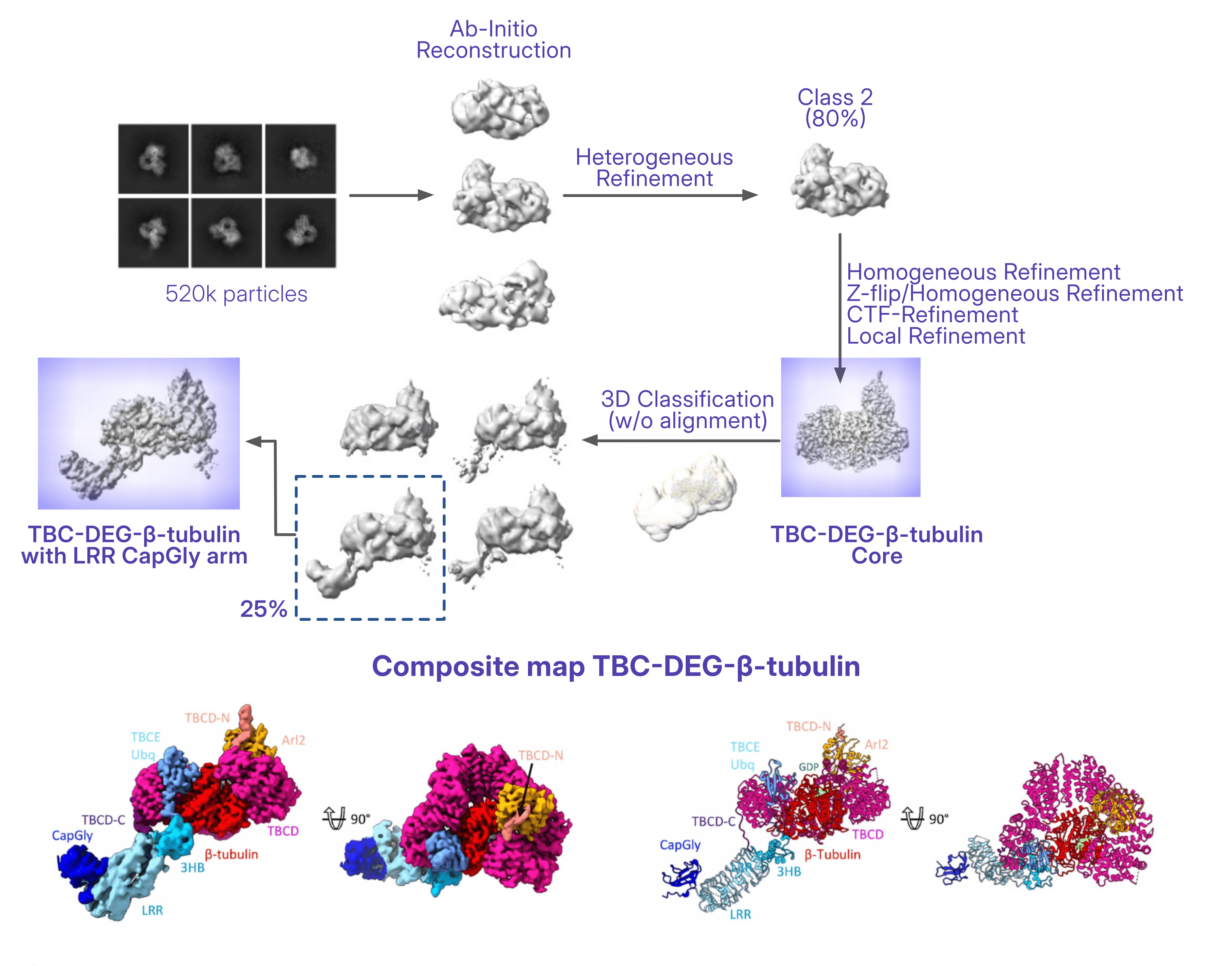
The non-crosslinked TBC-DEG–β-tubulin dataset was processed with multi-class Ab-Initio Reconstruction, followed by Heterogeneous Refinement to isolate the particles of interest. Subsequent Homogeneous Refinement, CTF Refinement, and Local Refinement produced a 3.6 Å reconstruction of the rigid TBC-DEG core, corresponding to the Arl2-bound β-tubulin intermediate that precedes α-tubulin incorporation. Because the TBCE LRR–CapGly arm remained flexible, 3D Classification without particle alignment was performed using focused masks encompassing the core and the expected position of the arm. Local refinement of this class resolved the full TBC-DEG–β-tubulin complex at lower resolution, and a composite map combining the high-resolution core with the refined TBCE arm was generated for model building.
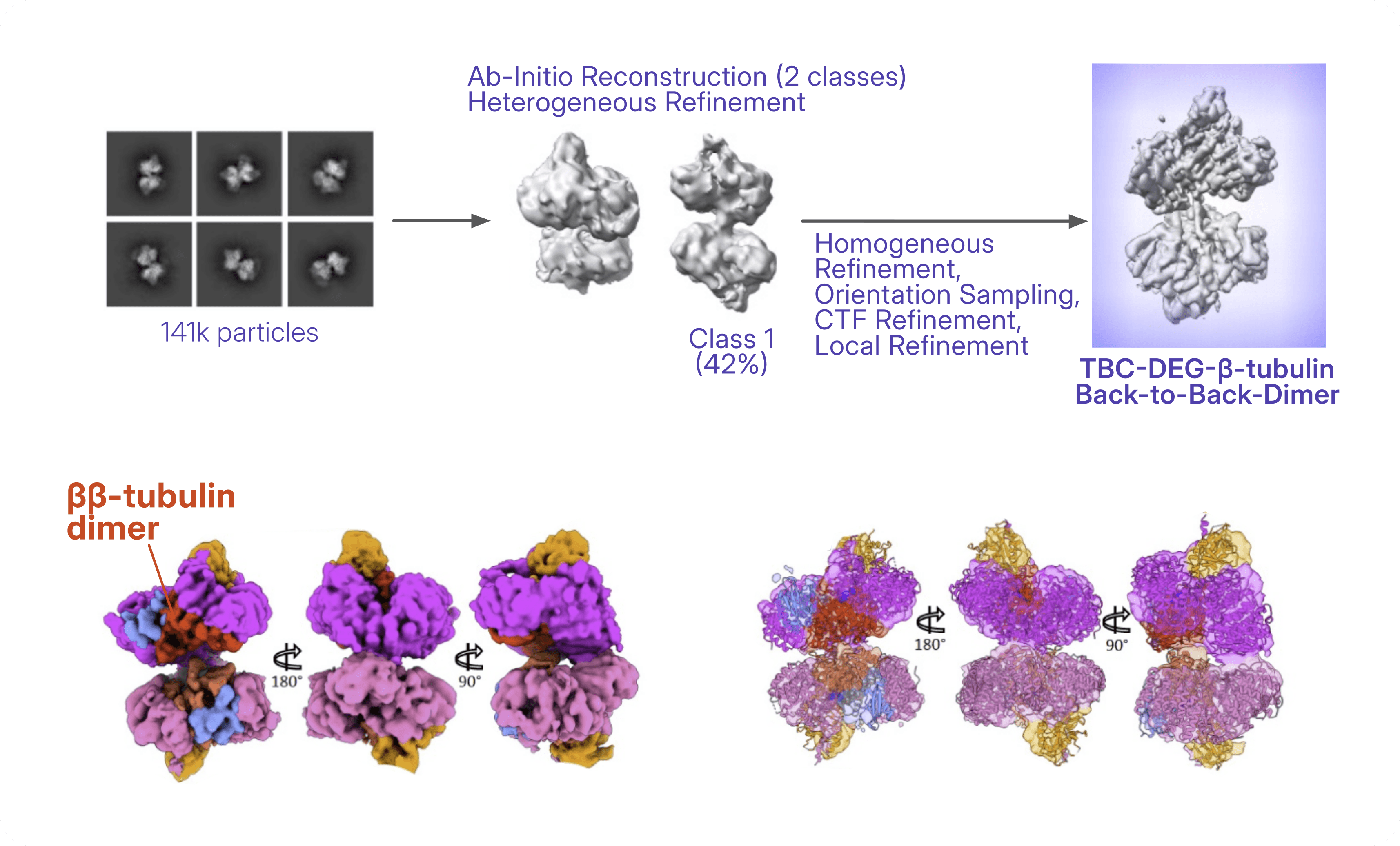
The crosslinked TBC-DEG–β-tubulin dataset was processed using multi-class Ab-Initio Reconstruction followed by Heterogeneous Refinement to isolate the rare back-to-back dimeric assembly stabilized by crosslinking. This structure captures a β-tubulin-bound dimeric state of the TBC-DEG complex, providing additional structural insight into higher-order assemblies that complement the monomeric intermediates used to reconstruct the overall catalytic mechanism.
A Structural Blueprint for Future Therapies
Rather than providing a single snapshot, the cryo-EM study adds several key β-tubulin-bound intermediates to an expanding structural framework.
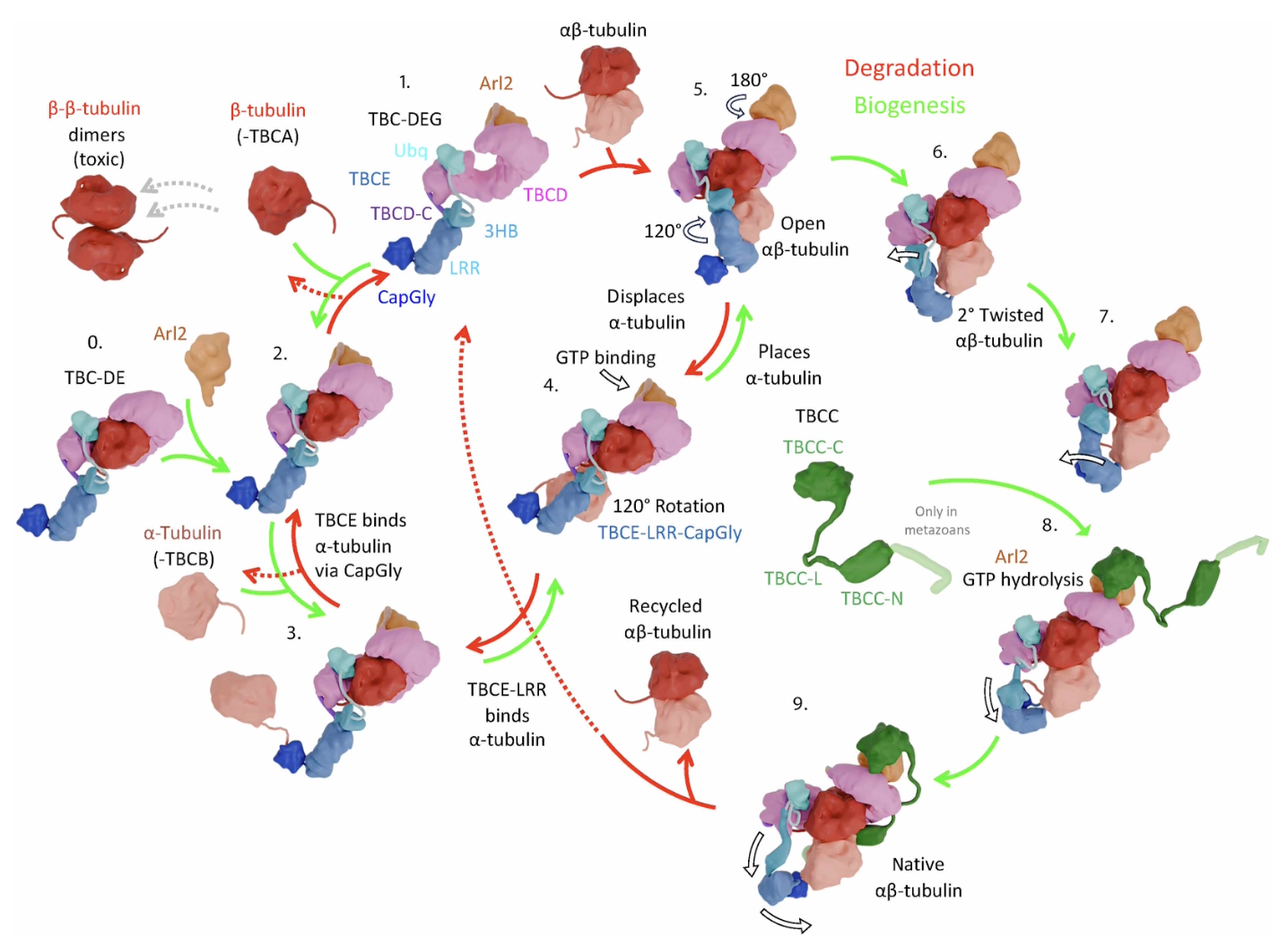
“There were two big surprises. The first was the nature of the catalytic transitions that place and displace α-tubulin: a lever-arm–like motion of the TBCE protein, triggered by GTP binding in Arl2 and transmitted over a long range through the TBCD scaffolding protein.”
- Dr. Jawdat Al-Bassam, Associate Professor, UCDavis (corresponding author)
The newly resolved structures reveal that TBC-DEG functions as a catalytic chaperone that actively remodels its conformation throughout the assembly cycle. They show how TBCD and TBCE stabilize β-tubulin, uncover a previously unknown nucleotide-free state of Arl2 associated with β-tubulin binding, and explain how Arl2-driven rearrangements position the flexible TBCE arm to rapidly recruit α-tubulin.
“The second [surprise] was the tail-to-tail dimerization of the cofactor–β-tubulin assemblies. This revealed a clear off-pathway homodimerization activity of β-tubulin that had not previously been understood. The cryo-EM class averages and 3D refined models showed just how prevalent these β–β-tubulin dimers were in the data. The interface mediating the β–β-tubulin dimers was the interface involved in binding α-tubulin, providing a crucial explanation for why this system maybe needed to correctly place α-tubulin beneath β-tubulin.”
- Dr. Jawdat Al-Bassam, Associate Professor, UCDavis (corresponding author)
Importantly, the authors also captured β-tubulin homodimeric assemblies, providing structural evidence for a competing off-pathway state that the catalytic cycle is proposed to prevent by efficiently channeling β-tubulin toward productive α/β heterodimer formation.
.](/images/blog/tubulin/back-to-back-dimers.png)
Together with previously determined α/β-tubulin complexes from one earlier publication by the same laboratory and two modeled transient states, these cryo-EM structures reveal a unified nine-step catalytic cycle explaining how the TBC-DEG machinery orchestrates the assembly and, when necessary, the disassembly of α/β-tubulin.
Beyond answering a long-standing question in cell biology, these findings provide a molecular framework for understanding how disease-causing mutations disrupt tubulin biogenesis. Mapping patient mutations onto these structures helps explain the origins of tubulin biogenesis disorders and provides a structural foundation for improving genetic diagnosis, interpreting newly discovered variants, and, in the longer term, guiding the development of therapies targeting this essential cellular machinery.
References
- Flex, E., Niceta, M., Cecchetti, S., Thiffault, I., Au, M. G., Capuano, A., ... & Tartaglia, M. (2016).Biallelic mutations in TBCD, encoding the tubulin folding cofactor D, perturb microtubule dynamics and cause early-onset encephalopathy. The American Journal of Human Genetics, 99(4), 962-973.
- Miyake, N., Fukai, R., Ohba, C., Chihara, T., Miura, M., Shimizu, H., ... & Matsumoto, N. (2016).Biallelic TBCD mutations cause early-onset neurodegenerative encephalopathy. The American Journal of Human Genetics, 99(4), 950-961.
- Martin, N., Jaubert, J., Gounon, P., Salido, E., Haase, G., Szatanik, M., & Guénet, J. L. (2002).A missense mutation in Tbce causes progressive motor neuronopathy in mice. Nature Genetics, 32(3), 443-447.
- Akhmanova, A., & Kapitein, L. C. (2022).Mechanisms of microtubule organization in differentiated animal cells. Nature Reviews Molecular Cell Biology, 23(8), 541-558.
- Binarová, P., & Tuszynski, J. (2019).Tubulin: structure, functions and roles in disease. Cells, 8(10), 1294.
- Al-Bassam, J. (2017). Revisiting the tubulin cofactors and Arl2 in the regulation of soluble αβ-tubulin pools and their effect on microtubule dynamics. Molecular Biology of the Cell, 28(3), 359-363.
- Taheri, A., Wang, Z., Singal, B. et al.Cryo-EM structures of the tubulin cofactors reveal the molecular basis of alpha/beta-tubulin biogenesis. Nat Commun17, 1405 (2026).
- Seong, Y., Kim, H., Byun, K., Park, Y. W., & Roh, S. H. (2025).Structural dissection of αβ-tubulin heterodimer assembly and disassembly by human tubulin–specific chaperones. Science, 390(6772), eady2708.