Malaria Invasion at Molecular Resolution: From Protein Inhibitors to Membrane Remodeling
Using single-particle cryo-EM, researchers revealed how the malaria parasite's moving junction remodels host membranes and used its native structure to guide the design of a protein inhibitor of parasite invasion.
Nearly half a million children under the age of five die from malaria every year [1]. The disease is caused by the parasite Plasmodium falciparum, whose complex life cycle alternates between humans and Anopheles mosquitoes. During this cycle, the parasite infects human liver cells (hepatocytes) and red blood cells (RBCs), as well as the salivary glands of mosquitoes, enabling transmission between hosts.
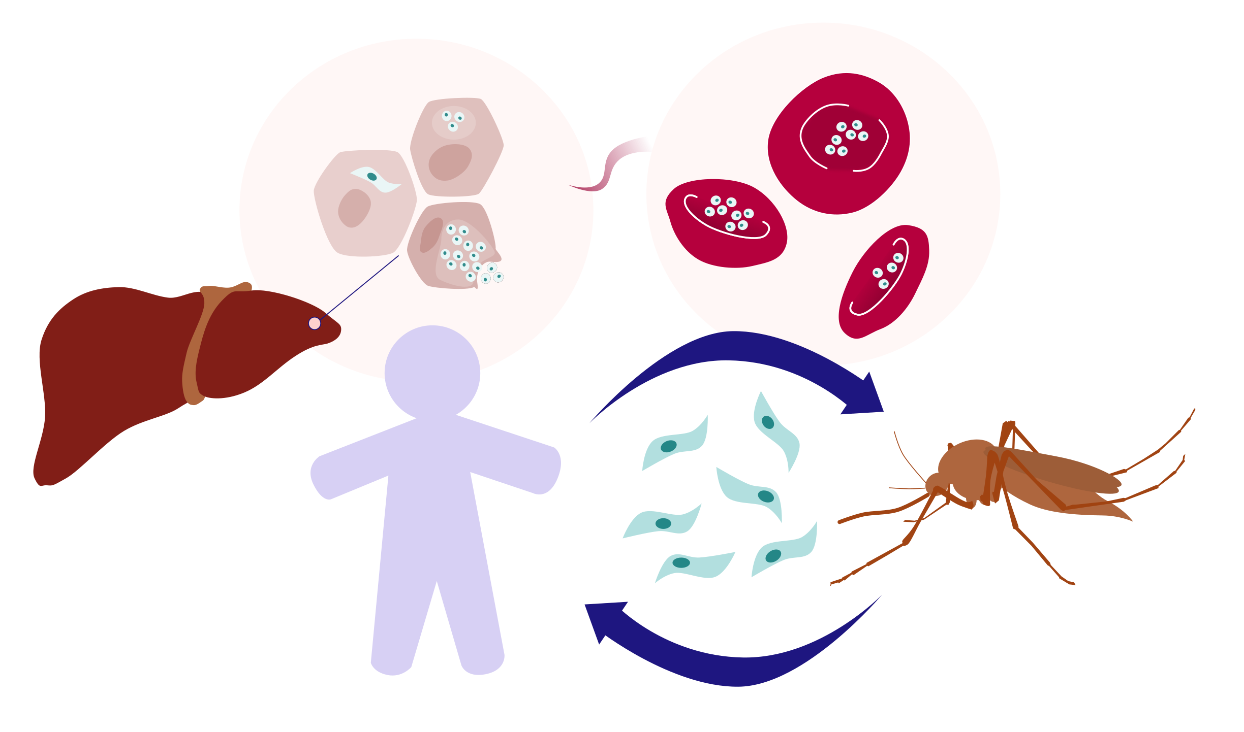
The parasite's life cycle has evolved to maximize transmission and relies on a sophisticated invasion mechanism. While the overall process has been modeled, many of its molecular details remain elusive. One of the earliest and most critical steps of invasion is the formation of a moving junction (MJ) between the parasite and the membrane of the target cell - whether a human red blood cell, a hepatocyte, or a mosquito salivary gland cell [2]. As the parasite actively penetrates the host cell, this ring-shaped junction acts both as an anchor that provides traction during invasion and as a molecular sieve that excludes host membrane proteins while allowing the parasite to enter.
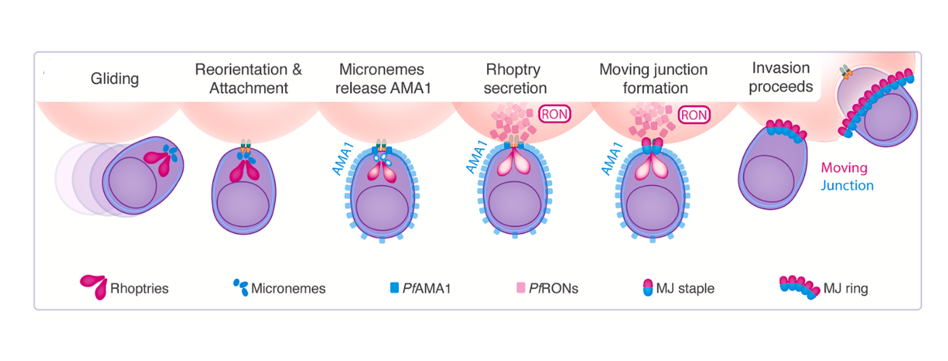 P. falciparum invasion of human RBCs, adapted from Figure 1 in Haile et al., 2026.
A recent study from Columbia University, published in Cell, presents the endogenous structure of the fundamental repeating unit of the moving junction (MJ staple), purified directly from invasion-stalled Plasmodium falciparum parasites. Using single-particle cryo-EM, the authors revealed a sailboat-shaped 1:1:1:1 assembly of apical membrane antigen 1 (PfAMA1) and rhoptry neck proteins 2, 4, and 5 (PfRON2, PfRON4, and PfRON5) at near-atomic resolution. This provides the most detailed mechanistic view to date of the moving junction unit and reveals a potential target for next-generation antimalarial therapeutics.
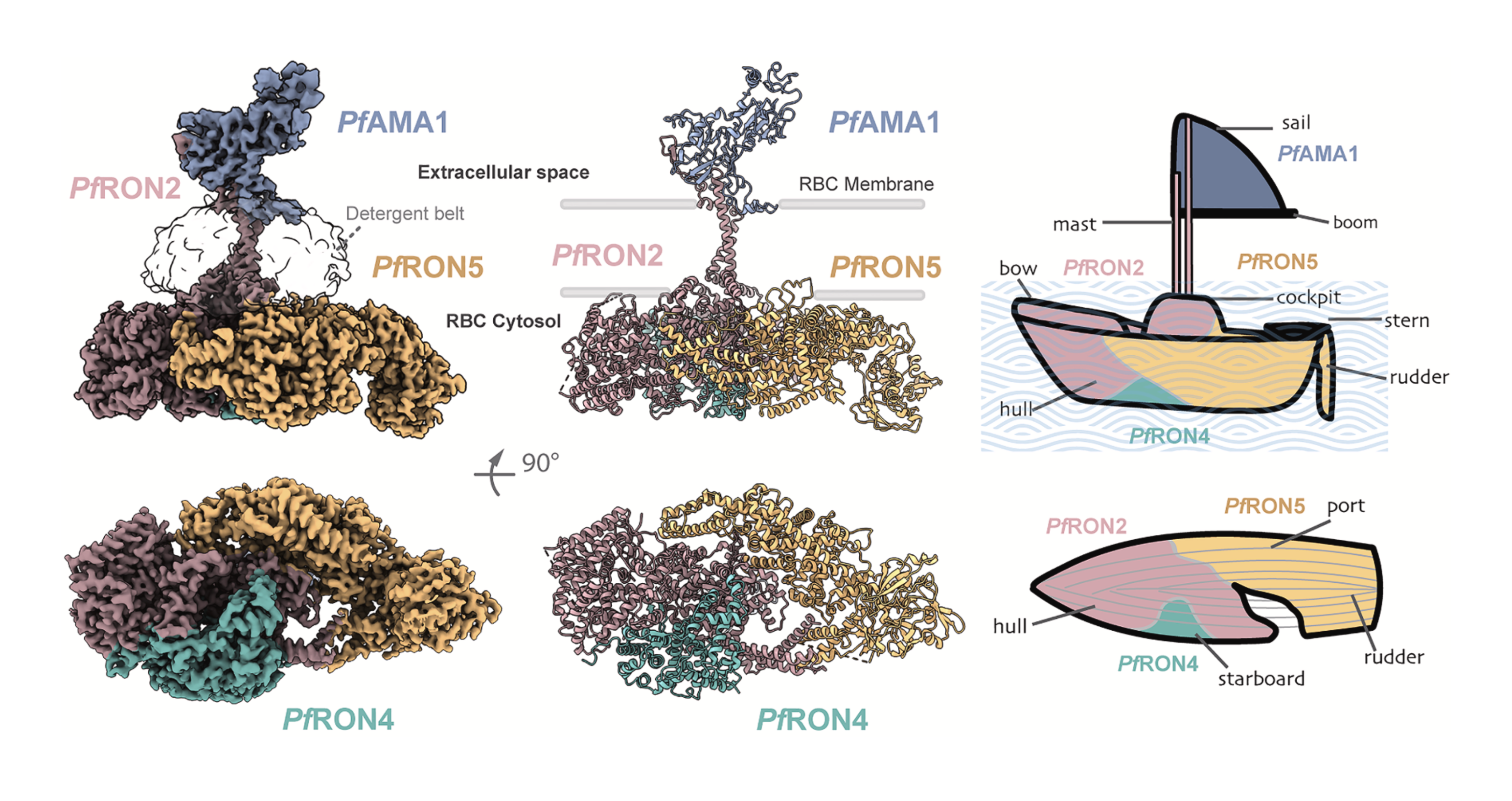 Cryo-EM map (left), atomic model (center), and schematic representation of the MJ staple. Adapted from Haile et al., 2026.
Cryo-EM Data Processing: Finding the Moving Junction in a Sea of Particles
The invasion step the authors aimed to capture exists for only 60 to 90 seconds in vivo. To ensure that the PfAMA1–PfRON2/4/5 assembly was faithfully captured, samples for single-particle cryo-EM were prepared using two different approaches. In the first, invasion was triggered by adding red blood cells to a highly synchronized P. falciparum culture and then stalled using cytochalasin D, allowing the natively assembled complex to be purified. In the second, the spontaneously assembled complex was isolated from non-stalled schizonts.
The two datasets were processed separately, and with only PfRON2 tagged for purification, the first challenge was figuring out which particles actually represented the moving junction complex.
"Since we enrich our samples directly from malaria parasites, and the proteins are not overexpressed at all, our micrographs always contain a heterogeneous mixture of native complexes that co-enrich with our complex. Figuring out which of the many different 2D class averages in the dataset were “ours” was quite challenging […]"
- Meseret Haile, Graduate Student in the Ho Lab, Columbia University (first author)
To identify the target particles and maximize their recovery, particularly across rare views, the authors adopted an iterative particle-picking strategy. Starting from ∼118,000 micrographs, the authors used Blob Picker and iterative 2D Classification to remove obvious junk particles.
Several additional rounds of 2D Classification were then used to identify promising classes. Selected particles were used for Ab-Initio Reconstruction, providing the first 3D maps of the complex. These initial reconstructions could then inform further particle picking, with new templates used to recover rare views with Template Picker and Topaz used to mine the micrographs for additional particles in parallel. Through this approach, the authors progressively expanded the particle set and its angular coverage before high-resolution refinement.
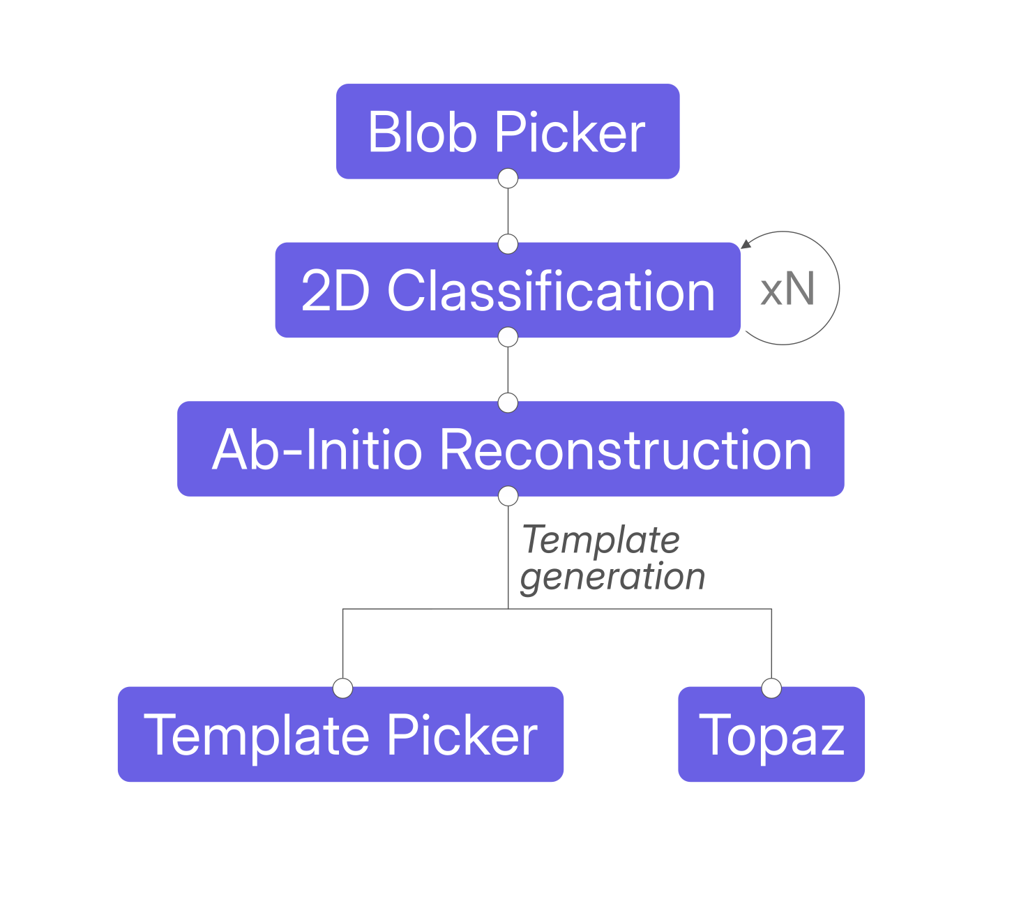 Data processing scheme adapted from Haile et al., 2026.
"[…] the moment when we finally identified the right set of 2D [classes] and they popped in Ab-Initio Refinement to reveal the beautiful and distinctive sailboat shape of our complex was probably the most memorable moment for us."
- Meseret Haile, Graduate Student in the Ho Lab, Columbia University (first author)
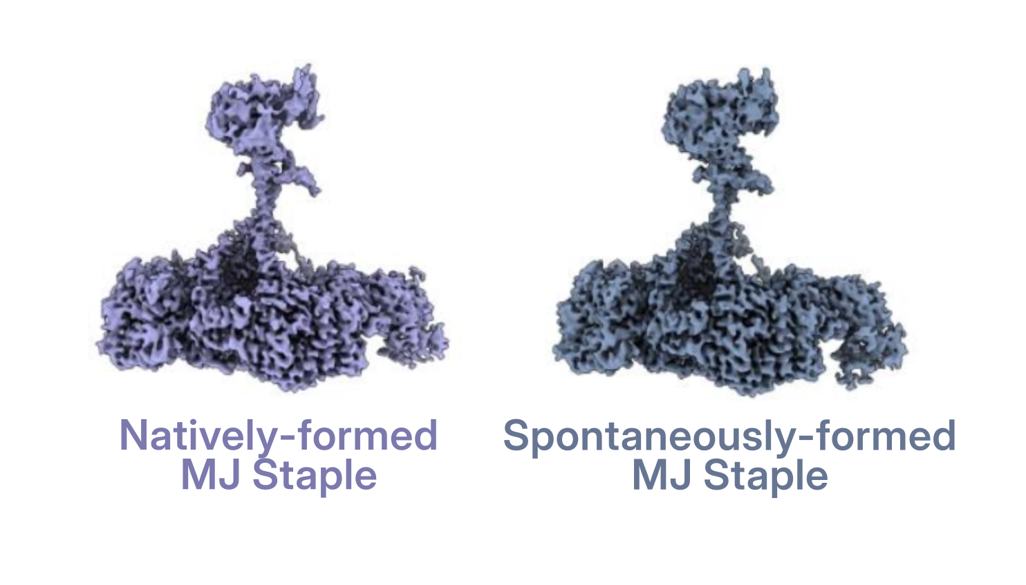 Adapted from Haile et al., 2026.
The resulting particle stacks from the invasion-stalled and from the spontaneously assembled moving junction complexes were refined independently using iterative Heterogeneous Refinement to further clean up the particle stack, followed by a final Homogeneous and Non-Uniform Refinement, producing two maps at 3.2 Å and 3.1 Å, respectively.
As the two structures proved essentially indistinguishable, the particle stacks were merged to obtain a 2.6 Å consensus reconstruction of the PfAMA1-RON2/4/5 complex. Because flexibility limited the local resolution of specific regions, focused processing strategies in CryoSPARC and RELION combining 3D Variability Analysis, particle subtraction, classification, and local refinement were used to improve the PfRON5 C-terminus and PfAMA1 regions before generating the final composite map.
The Native Structure of the MJ Staple Informs the de novo Inhibitory Protein Design
The sailboat-shaped complex revealed molecular details that were previously inaccessible. By providing the membrane context of all proteins involved in the MJ staple, the structure helps explain why the accessibility of previously studied antibodies depends on the stage of invasion.
Overall, the native cryo-EM structure shows that therapeutic accessibility depends not only on the binding epitope itself, but also on its spatial relationship with the parasite and host membranes. The authors used these structural constraints to guide a pilot study with BindCraft, designing potential protein inhibitors of complex formation. One candidate, A2, was experimentally tested, with in vitro studies confirming that it binds PfAMA1 at the PfRON2-binding cleft and blocks formation of the PfAMA1-PfRON2 complex.
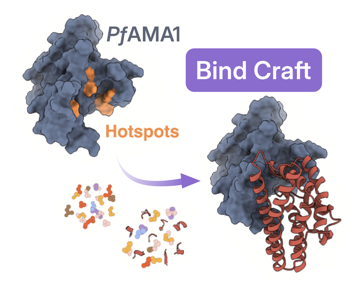 Adapted from Haile et al., 2026.
"I think our findings demonstrate that custom designed mini-protein inhibitors could be an exciting new strategy for antimalarial development and also highlight the power of having near-native structural information to guide the design of both conventional and next generation antimalarial therapies."
- Meseret Haile, Graduate Student in the Ho Lab, Columbia University (first author)
From Inhibitor Design to Biologic Functioning: Membrane Remodeling by the Moving Junction
The structure of PfAMA1–RON2/4/5 isolated in its native membrane context revealed another striking feature: PfRON2/4/5 displays several hallmarks of canonical membrane-remodeling proteins. The membrane-facing surface of the complex forms an unusually large, positively charged platform, which could strongly interact with the negatively charged inner leaflet of the RBC membrane. At its center, seven amphipathic helices from PfRON2 and PfRON5 insert into the membrane, with their hydrophobic faces buried deep within the lipid bilayer.
Rather than simply anchoring the parasite to the RBC, the moving junction may therefore actively help deform the host membrane as the parasite pushes its way inside.
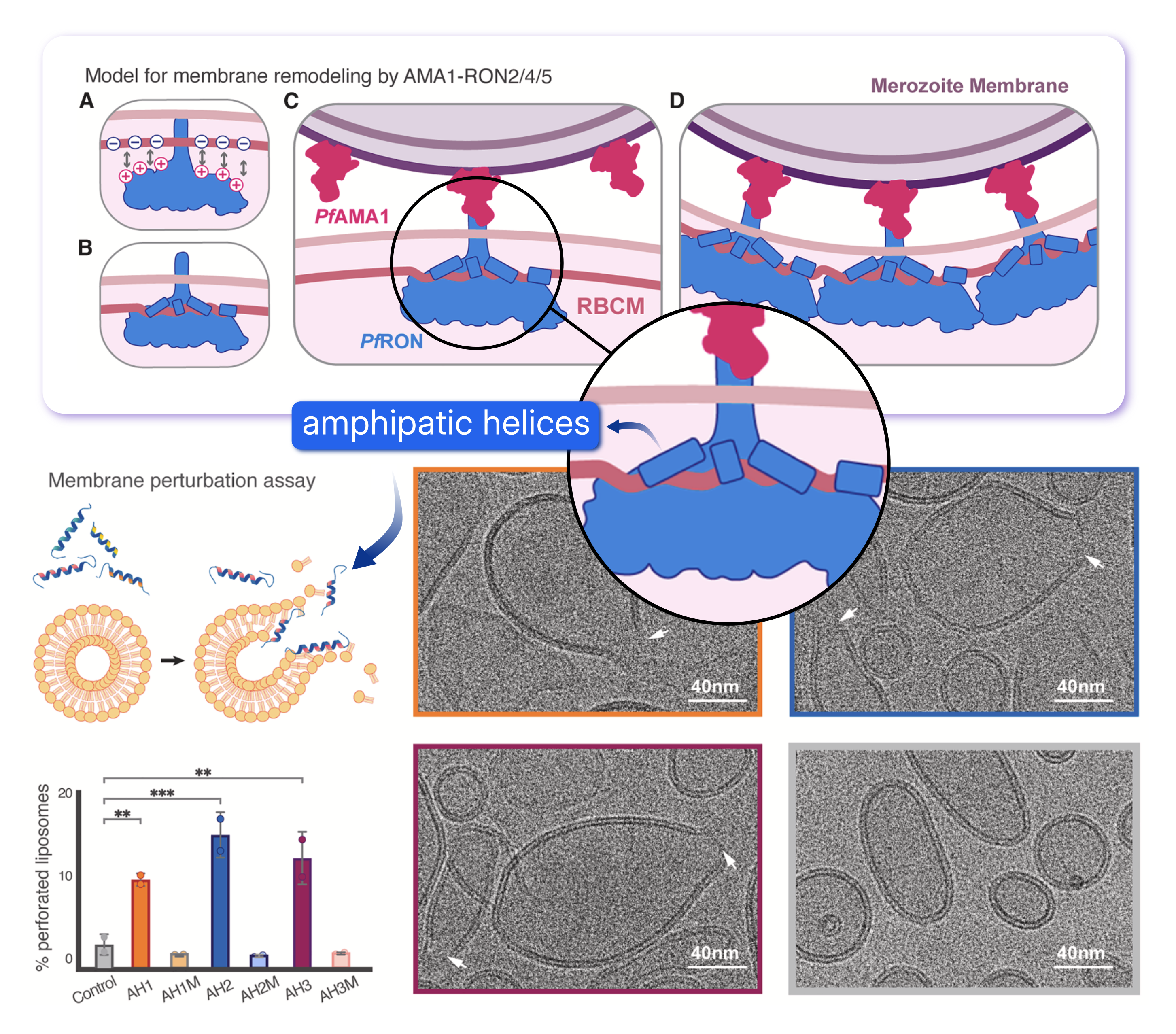 Top panel: proposed model of membrane remodelling from the MJ. Bottom panel: cryo-EM membrane perturbation assay. Adapted from Haile et al., 2026.
To experimentally test part of their model, the authors turned to cryo-EM once again. They synthesized peptides corresponding to the three PfRON2 amphipathic helices with the strongest hydrophobic character and incubated them with liposomes. Cryo-EM images revealed localized membrane thinning and pronounced perforations in the liposomes, while control liposomes remained intact. The more amphipathic the helix, the stronger the effect; when the helices were mutated to reduce their amphipathicity, membrane deformation was essentially lost. These experiments provide direct support for the idea that the PfRON2 amphipathic helices themselves can destabilize and remodel lipid membranes.
From cryo-EM sample preparation and the identification of the target complex among millions of particles across hundreds of thousands of micrographs, to de novo protein inhibitor design and a new model for membrane remodeling, this study shows how the effort invested in high-resolution structure determination can go far beyond the structure itself. By revealing the molecular details of parasite invasion, structural insights can generate new mechanistic hypotheses and provide a foundation for the development of next-generation antimalarial therapeutics.
References
- IHME, Global Burden of Disease (2025) – with major processing by Our World in Data. Child deaths from malaria.
- Besteiro, S., Dubremetz, J. F., & Lebrun, M. (2011).The moving junction of apicomplexan parasites: a key structure for invasion. Cellular Microbiology, 13(6), 797-805.